
Ultrasound scan

Preparing for an ultrasound scan
Before having some types of ultrasound scan, you may be asked to follow certain instructions to help improve the quality of the images produced.
For example, you may be advised to:
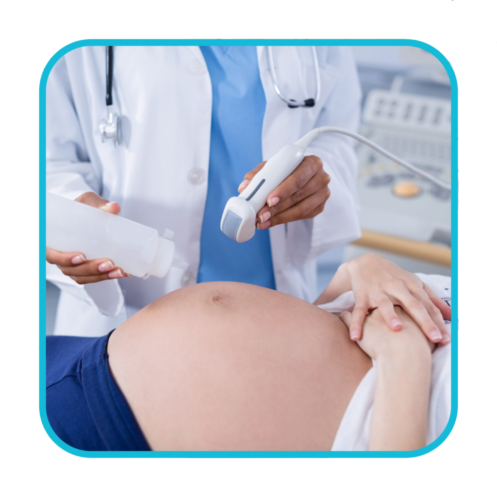
- drink water and not go to the toilet until after the scan – this may be needed before a scan of your unborn baby or your pelvic area
- avoid eating or drinking for several hours before the scan – this may be needed before a scan of your digestive system, including the liver and gallbladder
Depending on the area of your body being examined, the hospital may ask you to remove some clothing and wear a hospital gown.
What happens during an Ultrasound?
Most ultrasound scans last between 15 and 45 minutes. They usually take place in a hospital radiology department and are performed either by a doctor (radiologist) or a sonographer.
They can also be carried out in community locations such as GP practices, and may be performed by other healthcare professionals, such as midwives or physiotherapists who have been specially trained in ultrasound.
There are different kinds of ultrasound scans, depending on which part of the body is being scanned and why.
The 3 main types are:
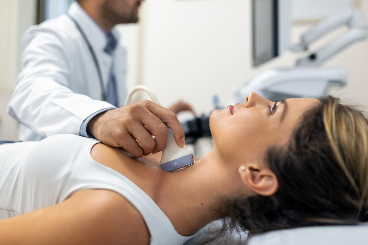
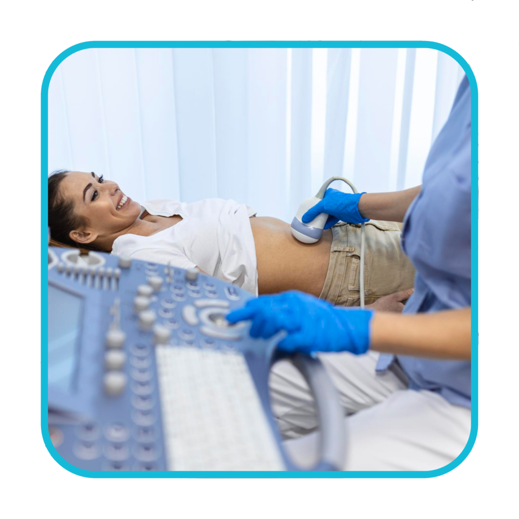
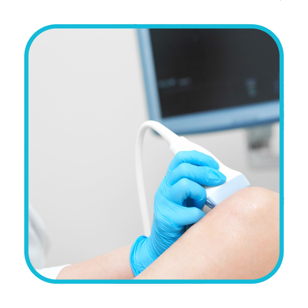
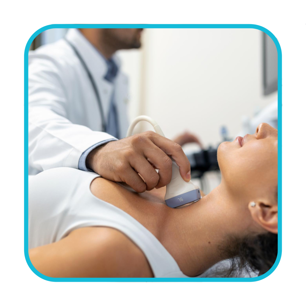
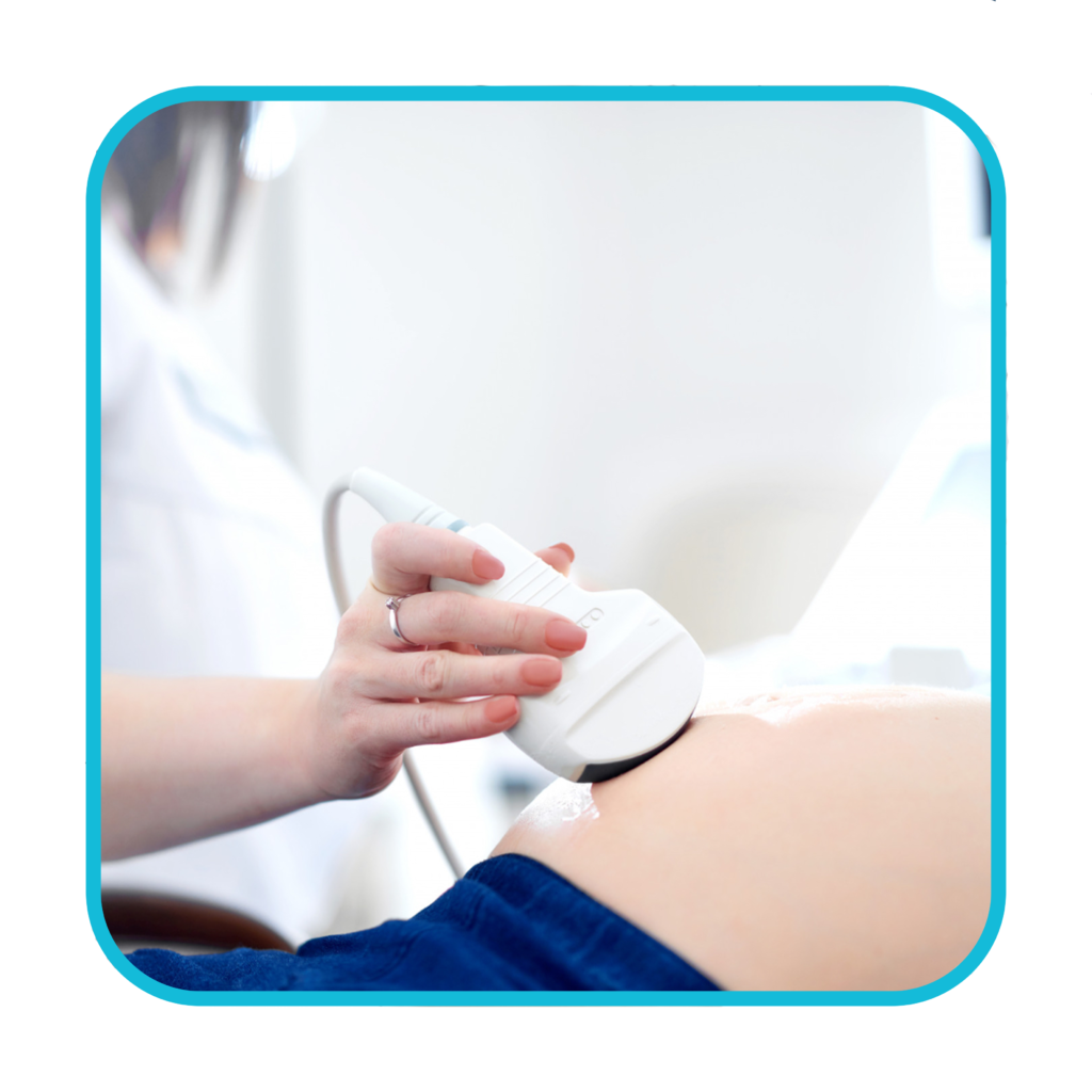
- external ultrasound scan – the probe is moved over the skin
- internal ultrasound scan – the probe is inserted into the body
- endoscopic ultrasound scan – the probe is attached to a long, thin, flexible tube (an endoscope) and passed further into the body – This type of scan is not undertaken at Greater Lancashire Hospital
Pricing
| Body part | Price |
|---|---|
| Abdominal aorta | 70 |
| Upper abdominal scan | 105 |
| Full abdominal scan | 150 |
| Female pelvic scan | 135 |
| Female pelvic including TV scan | 250 |
| Testes and Scrotal scan | 100 |
| KUB +/- prostate | 110 |
| Carotid scan | 250 |
| Wrist and hand scan | 175 |
| Knee scan one/two | 120/180 |
| Ankle and Foot scan | 175 |
| Hip scan one/two | 175/275 |
| Shoulder scan one/two | 175/275 |
| MSK soft tissue | 150 |
| Soft tissue + FNAC | 275 |
| US groin scan (hernia) | 200 |
| US neck/Thyroid scan | 150 |
| Thyroid + FNAC | 275 |
| Breast scanone/two | 125/175 |
| Breast + biopsy | 300 |
| DVTONE/TWO | 200/350 |
| Lower limbs arterial duplex | 250/400 |
| Upper limb arterial duplex | 250/400 |
| US varicose veins | 250/400 |
| Vein mapping | 200 |
| MSK injections | £150/- per injection |
Get in contact to book your appointment